
12-Week Ultrasound Explained
This ultrasound ensures everything is fine with you and baby, gives you some nice pictures, too.

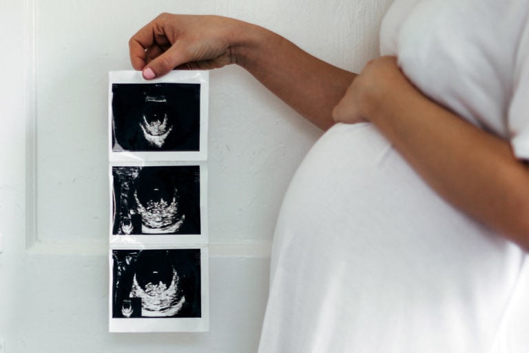
Around 12 weeks pregnant, you may have your first full-on anatomy scan.
What will happen during the 12-week ultrasound? Your ultrasound tech will put some (cold) gel on your belly and glide a transducer over it with mild pressure. The transducer sends out sound waves, and when those waves hit solid tissue, they create an image on the screen.
Tip: If you drink a cup or two of water before your scan, you’ll get a clearer image.
The 12-week ultrasound is not just for the pretty pictures. (Though those are the best part and make a great profile pic for your Babylist registry.) So why get it done? The scan ensures everything is fine with you and baby by:
Confirming the baby's heartbeat and your due date
Detecting potential problems
Making sure your body is ready for pregnancy
The tech looks at your fallopian tubes, uterus and placenta to see if you have a tilted uterus, placenta previa or other conditions that might make your pregnancy or labor unusual. These complications are very rare, but it's great to check them out early.
You won’t find out the sex for sure until the 20-week anatomy scan, but if you don’t want to know, be sure to tell your tech during your 12-week ultrasound.
You’ll also have the option of having a nuchal translucency scan, in which the tech measures the back of your baby’s neck to test for a few particular issues. According to Robyn Horsager-Boehrer, MD, Chief of Obstetrics and Gynecology at UT Southwestern Medical Center’s William P. Clements Jr. University Hospital, “The nuchal translucency represents the collection of fluid under the skin in the region of the posterior neck of the fetus. Increased size can be associated with an increased risk for chromosome abnormalities. This is most useful when combined with specific blood work. There are other congenital anomalies that are associated with increased measurements, especially cardiac abnormalities.”
Some possible conditions associated with a thicker-than-average nuchal area are Down syndrome, trisomy 18 or heart problems. This doesn’t mean your baby has any of these issues but has a greater risk of them. If this is the case, your doctor may suggest getting diagnostic tests such as Chorionic Villus (CVS) or amniocentesis.
In addition to your scan, your doctor will send you up for a blood screening test that measures plasma protein-A and human chorionic gonadotropin. “First-trimester screening with nuchal translucency and blood work assesses the risk of the three most common chromosome trisomies: Trisomy, 13, 18 and 21,” says Dr. Horsager-Boehrer. The results of the 12-week ultrasound along with your blood tests give your doctors an estimate of your risk. Statistically speaking this is all very unlikely, and your healthcare provider can provide all the relevant information.
“The [nuchal scan] results should give you an idea of what the likelihood is for those conditions and compare them to your risk before testing,” explains Dr. Horsager-Boehrer. “That estimate is based on your age. Once you have those results, you and your provider can discuss whether you want to pursue diagnostic testing (rather than screening) for chromosomal abnormalities.”
If your partner’s trying to figure out which prenatal visit to attend, this would be one of the most important—and most exciting—to join you for. It’s great to have a hand to hold and to coo over those 12-week ultrasound pictures together.